Home

steh učiť sa vzdialený umbilical sono kontrolór palica Vyjsť

Umbilical vein varix: Importance of ante- and post-natal monitoring by  ultrasound - ScienceDirect
Umbilical vein varix: Importance of ante- and post-natal monitoring by ultrasound - ScienceDirect

Portosystemic Shunt (Recanalized Umbilical Vein) - SONO.GALLERY

Jason H Collins MD,MSCR on Twitter: "@GEHealthcare Great,scan  Everything;the baby,the umbilical cord and the placenta then 3D print it  and give to mom. https://t.co/D0PdvAiN8G" / Twitter
Jason H Collins MD,MSCR on Twitter: "@GEHealthcare Great,scan Everything;the baby,the umbilical cord and the placenta then 3D print it and give to mom. https://t.co/D0PdvAiN8G" / Twitter
Ultrasound Video showing an Umbilical hernia. - YouTube

Ch. 25 Sono. Assessment of the Fetal Abdomen/Abdominal Wall Diagram |  Quizlet
Ch. 25 Sono. Assessment of the Fetal Abdomen/Abdominal Wall Diagram | Quizlet

Umbilical Artery Doppler – Sonographic Tendencies

Routine Identification of Placental Umbilical Cord Insertion Location  during Detailed Fetal Anatomic Ultrasound - Medical Update
Routine Identification of Placental Umbilical Cord Insertion Location during Detailed Fetal Anatomic Ultrasound - Medical Update

Portosystemic Shunt (Recanalized Umbilical Vein) - SONO.GALLERY

Portosystemic Shunt (Recanalized Umbilical Vein) - SONO.GALLERY

Normal appearance of the umbilicus by ultrasound, short axis. | Download  Scientific Diagram
Normal appearance of the umbilicus by ultrasound, short axis. | Download Scientific Diagram

Ultrasonography. A hypoechoic nodule of 3.9 × 2.3 × 1.9 cm in the... |  Download Scientific Diagram
Ultrasonography. A hypoechoic nodule of 3.9 × 2.3 × 1.9 cm in the... | Download Scientific Diagram

Abnormalities of the Umbilical Cord | IntechOpen

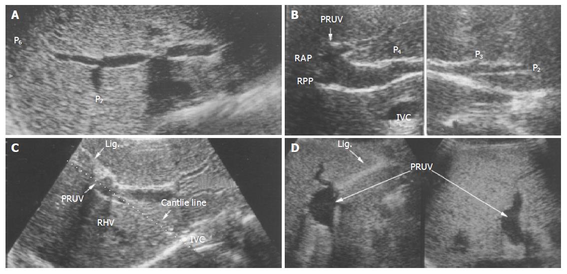
PDF] Are congenital malformations more frequent in fetuses with  intrahepatic persistent right umbilical vein? A comparative study. |  Semantic Scholar
PDF] Are congenital malformations more frequent in fetuses with intrahepatic persistent right umbilical vein? A comparative study. | Semantic Scholar

Differential diagnosis of umbilical polyps and granulomas in children:  sonographic and pathologic correlations
Differential diagnosis of umbilical polyps and granulomas in children: sonographic and pathologic correlations

Single umbilical artery | Radiology Reference Article | Radiopaedia.org

Differential diagnosis of umbilical polyps and granulomas in children:  sonographic and pathologic correlations
Differential diagnosis of umbilical polyps and granulomas in children: sonographic and pathologic correlations

Normal appearance of the umbilicus by ultrasound, short axis. | Download  Scientific Diagram
Normal appearance of the umbilicus by ultrasound, short axis. | Download Scientific Diagram

Median umbilical ligament - Wikipedia

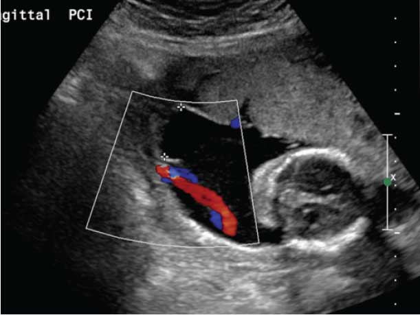
Umbilical vein varix: Importance of ante- and post-natal monitoring by  ultrasound - ScienceDirect
Umbilical vein varix: Importance of ante- and post-natal monitoring by ultrasound - ScienceDirect

Normal appearance of the umbilicus by ultrasound, short axis. | Download  Scientific Diagram
Normal appearance of the umbilicus by ultrasound, short axis. | Download Scientific Diagram

Small Bowel Obstruction Secondary to Strangulated Umbilical Hernia” in  “Strangulated Umbilical Hernia” on eScholarship
Small Bowel Obstruction Secondary to Strangulated Umbilical Hernia” in “Strangulated Umbilical Hernia” on eScholarship

Subhepatic course of the umbilical vein into intrahepatic part of the... |  Download Scientific Diagram
Subhepatic course of the umbilical vein into intrahepatic part of the... | Download Scientific Diagram

An anomaly in persistent right umbilical vein of portal vein diagnosed by  ultrasonography
An anomaly in persistent right umbilical vein of portal vein diagnosed by ultrasonography

Persistent Right Umbilical Vein

Single Umbilical Artery on Fetal Ultrasound - YouTube